Research (nuclear medicine physics)
Our research portfolio includes clinical trial support, independent research supported by NIHR and university funding as well as commercial research projects funded by industrial partners. Our key areas of expertise are radionuclide therapy and dosimetry, brain imaging and respiratory imaging.
Clinical trial support
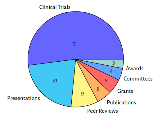
Our team provides medical physics expert advice in local and multi-centre clinical trials and commercial research studies including first in human studies. We set up approximately 20 new trials each year across nuclear medicine and PET. Our involvement includes developing and setting up imaging and therapy protocols, providing advice on image optimisation and dosimetry, and performing specialist quality assurance procedures for scanner accreditation and image quality harmonisation. Recent examples of clinical trials include the TRIPP_FFX radionuclide therapy study which uses P-32 microsphere therapy (Oncosil) for pancreatic cancer, and a range of neurology trials using amyloid and tau PET imaging to evaluate the efficacy of new disease modifying treatments for Alzheimer’s disease.
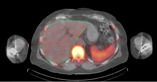
Radionuclide therapy and dosimetry
Radionuclide therapy dosimetry involves the calculation and measurement of radiation doses delivered to target tissues during therapeutic procedures, ensuring optimal treatment efficacy while minimizing radiation exposure to healthy organs. This precise assessment guides treatment planning and adjustments, enhancing patient safety and therapeutic outcomes.
Nuclear medicine physics at UHS have developed pragmatic, patient-specific dosimetry solutions for target organs and organs at risk in a range of therapeutic applications. We use these techniques clinically and as part of multi-centre clinical trials evaluating the safety and efficacy of MRT, as well as in commercial projects supporting industrial partners.
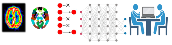
Quantitative analysis of radionuclide imaging of the brain
The group has an established record of designing, validating, and implementing a range of quantitative methods of analysing DaTSCAN datasets. With their high binding affinity for pre-synaptic dopamine transporters (DAT), these scans are able to diagnose Parkinson’s disease and offer a differential diagnosis over other neurological disorders with a similar clinical presentation. The group has experience of using regional uptake, shape, and pattern recognition to aid the diagnosis.
Cerebral perfusion imaging is currently attracting renewed interest, as a result of the possibility of drug treatment of Alzheimer’s disease. Imaging provides a valuable method of objectively assessing the response to treatment and also the possibility of aiding and confirming diagnosis. We have pioneered the routine clinical use of statistical parametric mapping (SPM) and other quantitative and highly sensitive approaches to image analysis of these scans, enabling response and diagnosis to be called with greater confidence.
As part of the BRAIN AI fellowship (Biomarker Research Assessing Inflammation in Neurodegeneration using AI) funded by the NIHR we have identified novel inflammation markers which offer complementary information to brain imaging, improving dementia prognosis. We have also developed a prototype AI system to support early diagnosis of dementia from brain perfusion scans.
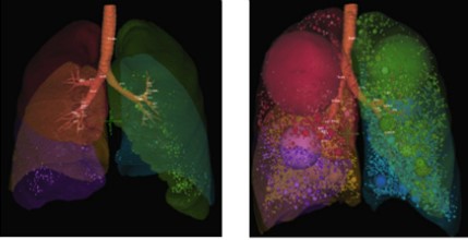
Respiratory imaging
A key area of our research is respiratory imaging and this is carried out jointly with the NIHR Respiratory Biomedical Research Centre at UHS.
Studying the pathway of inhaled radiolabelled aerosols in the body provides unique experimental data on the pattern of aerosol deposition within the lung. This research is valuable in optimising inhaled delivery of drugs for the treatment of asthma helping us understand the health effects of particulate pollution in the atmosphere.
Furthermore, measuring the biodistribution of antibody labelled radiopharmaceuticals in the lungs provides opportunities for new immunotherapy targets in chronic obstructive pulmonary disease.